| Title | Course Number | Author | Publisher | ISBN | Copyright | Who Owns it |
Website |
|---|
Living Bravely Courses # 8500 / 1 Allen C. Guelzo The Great Courses 156585763-1 2003 The History of the U.S. 2nd ediitioncover.jpg
Spain, France, and the Netherlands Courses # 8500 / 2 Allen C. Guelzo The Great Courses 156585763-1 2003 The History of the U.S. 2nd ediitioncover.jpg
Gentlemen in the Wilderness Courses # 8500 / 3 Allen C. Guelzo The Great Courses 156585763-1 2003 The History of the U.S. 2nd ediitioncover.jpg
Radicals in the Wilderness Courses # 8500 / 4 Allen C. Guelzo The Great Courses 156585763-1 2003 The History of the U.S. 2nd ediitioncover.jpg
Traders in the Wilderness Courses # 8500 / 5 Allen C. Guelzo The Great Courses 156585763-1 2003 The History of the U.S. 2nd ediitioncover.jpg
An Economy of Slaves Courses # 8500 / 6 Allen C. Guelzo The Great Courses 156585763-1 2003 The History of the U.S. 2nd ediitioncover.jpg
Printers, Painters, and Preachers Courses # 8500 / 7 Allen C. Guelzo The Great Courses 156585763-1 2003 The History of the U.S. 2nd ediitioncover.jpg
The Great Awakening Courses # 8500 / 8 Allen C. Guelzo The Great Courses 156585763-1 2003 The History of the U.S. 2nd ediitioncover.jpg
The Great War for Empire Courses # 8500 / 9 Allen C. Guelzo The Great Courses 156585763-1 2003 The History of the U.S. 2nd ediitioncover.jpg
The Rejection of Empire Courses # 8500 / 10 Allen C. Guelzo The Great Courses 156585763-1 2003 The History of the U.S. 2nd ediitioncover.jpg
The American Revolution- Politics aqnd People Courses # 8500 / 11 Allen C. Guelzo The Great Courses 156585763-1 2003 The History of the U.S. 2nd ediitioncover.jpg
The American Revolution- Howe's War Courses # 8500 / 12 Allen C. Guelzo The Great Courses 156585763-1 2003 The History of the U.S. 2nd ediitioncover.jpg
The American Revolution- Washington's War Courses # 8500 / 13 Allen C. Guelzo The Great Courses 156585763-1 2003 The History of the U.S. 2nd ediitioncover.jpg
Creating the Constitution Courses # 8500 / 14 Allen C. Guelzo The Great Courses 156585763-1 2003 The History of the U.S. 2nd ediitioncover.jpg
Hamilton's Republic Courses # 8500 / 15 Allen C. Guelzo The Great Courses 156585763-1 2003 The History of the U.S. 2nd ediitioncover.jpg
Republican and Federalists Courses # 8500 / 16 Allen C. Guelzo The Great Courses 156585763-1 2003 The History of the U.S. 2nd ediitioncover.jpg
Adams and Liberty Courses # 8500 / 17 Allen C. Guelzo The Great Courses 156585763-1 2003 The History of the U.S. 2nd ediitioncover.jpg
The Jefferson Reaction Courses # 8500 / 18 Allen C. Guelzo The Great Courses 156585763-1 2003 The History of the U.S. 2nd ediitioncover.jpg
Territory and Treason Courses # 8500 / 19 Allen C. Guelzo The Great Courses 156585763-1 2003 The History of the U.S. 2nd ediitioncover.jpg
The Agrarian Republic Courses # 8500 / 20 Allen C. Guelzo The Great Courses 156585763-1 2003 The History of the U.S. 2nd ediitioncover.jpg
The Disastrous War of 1812 Courses # 8500 / 21 Allen C. Guelzo The Great Courses 156585763-1 2003 The History of the U.S. 2nd ediitioncover.jpg
The" American System" Courses # 8500 / 22 Allen C. Guelzo The Great Courses 156585763-1 2003 The History of the U.S. 2nd ediitioncover.jpg
A Nation Announcing Itself Courses # 8500 / 23 Allen C. Guelzo The Great Courses 156585763-1 2003 The History of the U.S. 2nd ediitioncover.jpg
National Republican Follies Courses # 8500 / 24 Allen C. Guelzo The Great Courses 156585763-1 2003 The History of the U.S. 2nd ediitioncover.jpg
The Second Great Awakening Courses # 8500 / 25 Allen C. Guelzo The Great Courses 156585763-1 2003 The History of the U.S. 2nd ediitioncover.jpg
Dark Satanic Mills Courses # 8500 / 26 Allen C. Guelzo The Great Courses 156585763-1 2003 The History of the U.S. 2nd ediitioncover.jpg
The Politics of Distrust Courses # 8500 / 28 Allen C. Guelzo The Great Courses 156585763-1 2003 The History of the U.S. 2nd ediitioncover.jpg
The Monster Bank Courses # 8500 / 29 Allen C. Guelzo The Great Courses 156585763-1 2003 The History of the U.S. 2nd ediitioncover.jpg
Whigs and Democrats Courses # 8500 / 30 Allen C. Guelzo The Great Courses 156585763-1 2003 The History of the U.S. 2nd ediitioncover.jpg
American Romanticism Courese # 8500 / 31 Allen C. Guelzo The Great Courses 156585763-1 2003 The History of the U.S. 2nd ediitioncover.jpg
The Age of Reform Courses # 8500 / 32 Allen C. Guelzo The Great Courses 156585763-1 2003 The History of the U.S. 2nd ediitioncover.jpg
Southern Society and the Defense of Slavery Courses # 8500 / 33 Allen C. Guelzo The Great Courses 156585763-1 2003 The History of the U.S. 2nd ediitioncover.jpg
Whose Manifest Destiny? Courses # 8500 / 34 Allen C. Guelzo The Great Courses 156585763-1 2003 The History of the U.S. 2nd ediitioncover.jpg
The Mexican War Courses # 8500 / 35 Allen C. Guelzo The Great Courses 156585763-1 2003 The History of the U.S. 2nd ediitioncover.jpg
The Great Compromise Courses # 8500 / 36 Allen C. Guelzo The Great Courses 156585763-1 2003 The History of the U.S. 2nd ediitioncover.jpg
Sectional Tenisons Escalate Courses # 8500 / 37 Allen C. Guelzo The Great Courses 156585763-1 2003 The History of the U.S. 2nd ediitioncover.jpg
Drifting Toward Disaster Courses # 8500 / 38 Allen C. Guelzo The Great Courses 156585763-1 2003 The History of the U.S. 2nd ediitioncover.jpg
The Coming of War Courses # 8500 / 39 Allen C. Guelzo The Great Courses 156585763-1 2003 The History of the U.S. 2nd ediitioncover.jpg
The First Year of Fighting Courses # 8500 / 40 Allen C. Guelzo The Great Courses 156585763-1 2003 The History of the U.S. 2nd ediitioncover.jpg
Shifting Tides of Battle Courses # 8500 / 41 Allen C. Guelzo The Great Courses 156585763-1 2003 The History of the U.S. 2nd ediitioncover.jpg
Diplomatic Clashes and Sustaining the War Courses # 8500 / 42 Allen C. Guelzo The Great Courses 156585763-1 2003 The History of the U.S. 2nd ediitioncover.jpg
Behindthe Lines-Politics and Economies Courses # 8500 / 43 Allen C. Guelzo The Great Courses 156585763-1 2003 The History of the U.S. 2nd ediitioncover.jpg
African Americans in Wartime Courses # 8500 / 44 Allen C. Guelzo The Great Courses 156585763-1 2003 The History of the U.S. 2nd ediitioncover.jpg
The Union Drive to Victory Courses # 8500 / 45 Allen C. Guelzo The Great Courses 156585763-1 2003 The History of the U.S. 2nd ediitioncover.jpg
Presidential Reconstruction Courses # 8500 / 46 Allen C. Guelzo The Great Courses 156585763-1 2003 The History of the U.S. 2nd ediitioncover.jpg
Congress Takes Command Courses # 8500 / 47 Allen C. Guelzo The Great Courses 156585763-1 2003 The History of the U.S. 2nd ediitioncover.jpg
Reconstruction Ends Courses # 8500 / 48 Allen C. Guelzo The Great Courses 156585763-1 2003 The History of the U.S. 2nd ediitioncover.jpg
Industrialization Courses # 8500 / 49 Allen C. Guelzo The Great Courses 156585763-1 2003 The History of the U.S. 2nd ediitioncover.jpg
Transcontinental Railroads Courses # 8500 / 50 Allen C. Guelzo The Great Courses 156585763-1 2003 The History of the U.S. 2nd ediitioncover.jpg
The Last Indian War Courses # 8500 / 51 Allen C. Guelzo The Great Courses 156585763-1 2003 The History of the U.S. 2nd ediitioncover.jpg
Farming the Great Plains Courses # 8500 / 52 Allen C. Guelzo The Great Courses 156585763-1 2003 The History of the U.S. 2nd ediitioncover.jpg
African Americans after Reconstruction Courses # 8500 / 53 Allen C. Guelzo The Great Courses 156585763-1 2003 The History of the U.S. 2nd ediitioncover.jpg
Men and Women Courses # 8500 / 54 Allen C. Guelzo The Great Courses 156585763-1 2003 The History of the U.S. 2nd ediitioncover.jpg
Religion in Victorian America Courses # 8500 / 55 Allen C. Guelzo The Great Courses 156585763-1 2003 The History of the U.S. 2nd ediitioncover.jpg
The Populists Courses # 8500 / 56 Allen C. Guelzo The Great Courses 156585763-1 2003 The History of the U.S. 2nd ediitioncover.jpg
The New Immigration Courses # 8500 / 57 Allen C. Guelzo The Great Courses 156585763-1 2003 The History of the U.S. 2nd ediitioncover.jpg
City Life Courses # 8500 / 58 Allen C. Guelzo The Great Courses 156585763-1 2003 The History of the U.S. 2nd ediitioncover.jpg
Labor and Capital Courses # 8500 / 59 Allen C. Guelzo The Great Courses 156585763-1 2003 The History of the U.S. 2nd ediitioncover.jpg
Theodore Roosevelt and Progressivism Courses # 8500 / 60 Allen C. Guelzo The Great Courses 156585763-1 2003 The History of the U.S. 2nd ediitioncover.jpg
Mass Production Courses # 8500 / 61 Allen C. Guelzo The Great Courses 156585763-1 2003 The History of the U.S. 2nd ediitioncover.jpg
World War I - The Road to intervention Courses # 8500 / 62 Allen C. Guelzo The Great Courses 156585763-1 2003 The History of the U.S. 2nd ediitioncover.jpg
World War I - Versailles and Wilson's Gambit Courses # 8500 / 63 Allen C. Guelzo The Great Courses 156585763-1 2003 The History of the U.S. 2nd ediitioncover.jpg
The 1920's Courses # 8500 / 64 Allen C. Guelzo The Great Courses 156585763-1 2003 The History of the U.S. 2nd ediitioncover.jpg
The Wall Street Crash and the Great Depression Courses # 8500 / 65 Allen C. Guelzo The Great Courses 156585763-1 2003 The History of the U.S. 2nd ediitioncover.jpg
The New Deal Courses # 8500 / 66 Allen C. Guelzo The Great Courses 156585763-1 2003 The History of the U.S. 2nd ediitioncover.jpg
World War II - The Road to Pearl Harbor Courses # 8500 / 67 Allen C. Guelzo The Great Courses 156585763-1 2003 The History of the U.S. 2nd ediitioncover.jpg
World War II - The European Theater Courses # 8500 / 68 Allen C. Guelzo The Great Courses 156585763-1 2003 The History of the U.S. 2nd ediitioncover.jpg
World War II - The Pacific Theater Courses # 8500 / 69 Allen C. Guelzo The Great Courses 156585763-1 2003 The History of the U.S. 2nd ediitioncover.jpg
The Cold War Courses # 8500 / 70 Allen C. Guelzo The Great Courses 156585763-1 2003 The History of the U.S. 2nd ediitioncover.jpg
The Korean War and McCarthyism Courses # 8500 / 71 Allen C. Guelzo The Great Courses 156585763-1 2003 The History of the U.S. 2nd ediitioncover.jpg
The Affluent Society Courses # 8500 / 72 Allen C. Guelzo The Great Courses 156585763-1 2003 The History of the U.S. 2nd ediitioncover.jpg
The Civil Rights Movement Courses # 8500 / 73 Allen C. Guelzo The Great Courses 156585763-1 2003 The History of the U.S. 2nd ediitioncover.jpg
The New Frontier and the Great Society Courses # 8500 / 74 Allen C. Guelzo The Great Courses 156585763-1 2003 The History of the U.S. 2nd ediitioncover.jpg
The Rise of Mass Media Courses # 8500 / 75 Allen C. Guelzo The Great Courses 156585763-1 2003 The History of the U.S. 2nd ediitioncover.jpg
The Vietnam War Courses # 8500 / 76 Allen C. Guelzo The Great Courses 156585763-1 2003 The History of the U.S. 2nd ediitioncover.jpg
The Women's Movement Courses # 8500 / 77 Allen C. Guelzo The Great Courses 156585763-1 2003 The History of the U.S. 2nd ediitioncover.jpg
Nixon and Watergate Courses # 8500 / 78 Allen C. Guelzo The Great Courses 156585763-1 2003 The History of the U.S. 2nd ediitioncover.jpg
Enviromentalism Courses # 8500 / 79 Allen C. Guelzo The Great Courses 156585763-1 2003 The History of the U.S. 2nd ediitioncover.jpg
Religion in Twentieth -Century America Courses # 8500 / 80 Allen C. Guelzo The Great Courses 156585763-1 2003 The History of the U.S. 2nd ediitioncover.jpg
Carter and the Reagan Revolution Courses # 8500 / 81 Allen C. Guelzo The Great Courses 156585763-1 2003 The History of the U.S. 2nd ediitioncover.jpg
The New World Order Courses # 8500 / 82 Allen C. Guelzo The Great Courses 156585763-1 2003 The History of the U.S. 2nd ediitioncover.jpg
Clinton's America and the Millennium Courses # 8500 / 83 Allen C. Guelzo The Great Courses 156585763-1 2003 The History of the U.S. 2nd ediitioncover.jpg
Reflections Courses # 8500 / 84 Allen C. Guelzo The Great Courses 156585763-1 2003 The History of the U.S. 2nd ediitioncover.jpg
America—The Philosophical Experiment Courses # 4820 / 1 Joseph F. Kobylka The Great Courses 159803264-X 2006 CyclesofAmericanPoliticalThoughtcover.jpg
Historical Baggage Courses # 4820 / 2 Joseph F. Kobylka The Great Courses 159803264-X CyclesofAmericanPoliticalThoughtcover.jpg
Theorectial Baggage Courses # 4820 / 3 Joseph F. Kobylka The Great Courses 159803264-X CyclesofAmericanPoliticalThoughtcover.jpg
A Purtian Beginning Courses # 4820 / 4 Joseph F. Kobylka The Great Courses 159803264-X CyclesofAmericanPoliticalThoughtcover.jpg
Expansion and Individualism Courses # 4820 / 5 Joseph F. Kobylka The Great Courses 159803264-X CyclesofAmericanPoliticalThoughtcover.jpg
The Revolutionary Context Courses # 4820 / 6 Joseph F. Kobylka The Great Courses 159803264-X CyclesofAmericanPoliticalThoughtcover.jpg
The Road to the Declaration of Independence Courses # 4820 / 7 Joseph F. Kobylka The Great Courses 159803264-X CyclesofAmericanPoliticalThoughtcover.jpg
A "Natural" Revolutionary—Thomas Paine Courses # 4820 / 8 Joseph F. Kobylka The Great Courses 159803264-X CyclesofAmericanPoliticalThoughtcover.jpg
The Unconscious Dialectic of Crèvecoeur Courses # 4820 / 9 Joseph F. Kobylka The Great Courses 159803264-X CyclesofAmericanPoliticalThoughtcover.jpg
John Adams—"Constitutionalist" Courses # 4820 / 10 Joseph F. Kobylka The Great Courses 159803264-X CyclesofAmericanPoliticalThoughtcover.jpg
A Political Constitution Courses # 4820 / 11 Joseph F. Kobylka The Great Courses 159803264-X CyclesofAmericanPoliticalThoughtcover.jpg
A Philosophical Constitution—Faction Courses # 4820 / 12 Joseph F. Kobylka The Great Courses 159803264-X CyclesofAmericanPoliticalThoughtcover.jpg
3A Philosophical Constitution—Structure Courses # 4820 / 13 Joseph F. Kobylka The Great Courses 159803264-X CyclesofAmericanPoliticalThoughtcover.jpg
A Philosophical Constitution—Interpretation Courses # 4820 / 14 Joseph F. Kobylka The Great Courses 159803264-X CyclesofAmericanPoliticalThoughtcover.jpg
Disorganized Losers—The Anti-Federalists Courses # 4820 / 15 Joseph F. Kobylka The Great Courses 159803264-X CyclesofAmericanPoliticalThoughtcover.jpg
The "Genius" of Thomas Jefferson Courses # 4820 / 16 Joseph F. Kobylka The Great Courses 159803264-X CyclesofAmericanPoliticalThoughtcover.jpg
Jacksonian Democracy—The "People" Extended Courses # 4820 / 17 Joseph F. Kobylka The Great Courses 159803264-X CyclesofAmericanPoliticalThoughtcover.jpg
Iconoclastic Individualism—Thoreau Courses # 4820 / 18 Joseph F. Kobylka The Great Courses 159803264-X CyclesofAmericanPoliticalThoughtcover.jpg
Inclusionist Stirrings—Douglass and Stanton Courses # 4820 / 19 Joseph F. Kobylka The Great Courses 159803264-X CyclesofAmericanPoliticalThoughtcover.jpg
The Organic Socialism of Brownson Courses # 4820 / 20 Joseph F. Kobylka The Great Courses 159803264-X CyclesofAmericanPoliticalThoughtcover.jpg
American Feudalism—The Vision of Fitzhugh Courses # 4820 / 21 Joseph F. Kobylka The Great Courses 159803264-X CyclesofAmericanPoliticalThoughtcover.jpg
Constitutionalizing the Slave Class Courses # 4820 / 22 Joseph F. Kobylka The Great Courses 159803264-X CyclesofAmericanPoliticalThoughtcover.jpg
Lincoln's Reconstitution of America Courses # 4820 / 23 Joseph F. Kobylka The Great Courses 159803264-X CyclesofAmericanPoliticalThoughtcover.jpg
Equality in the Law and in Practice Courses # 4820 / 24 Joseph F. Kobylka The Great Courses 159803264-X CyclesofAmericanPoliticalThoughtcover.jpg
Social Darwinism and Economic Laissez-Faire Courses # 4820 / 25 Joseph F. Kobylka The Great Courses 159803264-X CyclesofAmericanPoliticalThoughtcover.jpg
Looking Backward, Looking Forward Courses #4820 / 26 Joseph F. Kobylka The Great Courses 159803264-X CyclesofAmericanPoliticalThoughtcover.jpg
Teddy Roosevelt and Progressivism Courses # 4820 / 27 Joseph F. Kobylka The Great Courses 159803264-X CyclesofAmericanPoliticalThoughtcover.jpg
Supreme Court and Laissez-Faire Courses # 4820 / 28 Joseph F. Kobylka The Great Courses 159803264-X CyclesofAmericanPoliticalThoughtcover.jpg
The Women's Movement and the 19th Amendment Courses # 4820 / 29 Joseph F. Kobylka The Great Courses 159803264-X CyclesofAmericanPoliticalThoughtcover.jpg
Eugene V. Debs and Working-Class Socialism Courses # 4820 / 30 Joseph F. Kobylka The Great Courses 159803264-X CyclesofAmericanPoliticalThoughtcover.jpg
Hamiltonian Means for Jeffersonian Ends Courses # 4820 / 31 Joseph F. Kobylka The Great Courses 159803264-X CyclesofAmericanPoliticalThoughtcover.jpg
FDR, the New Deal, and the Supreme Court Courses # 4820 / 32 Joseph F. Kobylka The Great Courses 159803264-X CyclesofAmericanPoliticalThoughtcover.jpg
The Racial Revolution Courses # 4820 / 33 Joseph F. Kobylka The Great Courses 159803264-X CyclesofAmericanPoliticalThoughtcover.jpg
The New Egalitarianism and Freedom Courses # 4820 / 34 Joseph F. Kobylka The Great Courses 159803264-X CyclesofAmericanPoliticalThoughtcover.jpg
The Reagan Revolution Courses # 4820 / 35 Joseph F. Kobylka The Great Courses 159803264-X CyclesofAmericanPoliticalThoughtcover.jpg
Cycles of American Political Conversations Courses # 4820 / 36 Joseph F. Kobylka The Great Courses 159803264-X CyclesofAmericanPoliticalThoughtcover.jpg
What Are Civil Liberties Courses # 8530 / 1 Professor John E. Finn The Great Courses 159803197-X civillibertiescover.jpg
The Bill of Rights- An Overview Courses # 8530 / 2 Professor John E. Finn The Great Courses 159803197-X civillibertiescover.jpg
Two Types of Liberties-Positive and Negative Courses # 8530 / 3 Professor John E. Finn The Great Courses 159803197-X civillibertiescover.jpg
The Court and Constitution Interpretation Courses # 8530 / 4 Professor John E. Finn The Great Courses 159803197-X civillibertiescover.jpg
Marbury v. Madison and Judicial Review Courses # 8530 / 5 Professor John E. Finn The Great Courses 159803197-X civillibertiescover.jpg
Private Property and the Founding Courses # 8530 / 6 Professor John E. Finn The Great Courses 159803197-X civillibertiescover.jpg
Lochner v. New York and Economic Due Process Courses # 8530 / 7 Professor John E. Finn The Great Courses 159803197-X civillibertiescover.jpg
The Takings Clause of the Fifth Amendment Courses # 8530 / 8 Professor John E. Finn The Great Courses 159803197-X civillibertiescover.jpg
Fundamental Rights - Privacy and Personhood Courses # 8530 / 9 Professor John E. Finn The Great Courses 159803197-X civillibertiescover.jpg
Privacy - Early Cases Courses # 8530 / 10 Professor John E. Finn The Great Courses 159803197-X civillibertiescover.jpg
Roe v. Wade and Reproductive Autonomy Courses # 8530 / 11 Professor John E. Finn The Great Courses 159803197-X civillibertiescover.jpg
Privacy and Autonomy - From Roe to Casey Courses # 8530 / 12 Professor John E. Finn The Great Courses 159803197-X civillibertiescover.jpg
Other Privacy Interests—Family Courses # 8530 / 13 Professor John E. Finn The Great Courses 159803197-X civillibertiescover.jpg
Other Privacy Interests—Sexuality Courses # 8530 / 14 Professor John E. Finn The Great Courses 159803197-X civillibertiescover.jpg
Same-Sex Marriages and the Constitution Courses # 8530 / 15 Professor John E. Finn The Great Courses 159803197-X civillibertiescover.jpg
The Right to Die and the Constitution Courses # 8530 / 16 Professor John E. Finn The Great Courses 159803197-X civillibertiescover.jpg
Cruel and Unusual? The Death Penalty Courses # 8530 / 17 Professor John E. Finn The Great Courses 159803197-X civillibertiescover.jpg
The First Amendment—An Overview Courses # 8530 / 18 Professor John E. Finn The Great Courses 159803197-X civillibertiescover.jpg
Internal Security and the First Amendment Courses # 8530 / 19 Professor John E. Finn The Great Courses 159803197-X civillibertiescover.jpg
Symbolic Speech and Expressive Conduct Courses # 8530 / 20 Professor John E. Finn The Great Courses 159803197-X civillibertiescover.jpg
Indecency and Obscenity Courses # 8530 / 21 Professor John E. Finn The Great Courses 159803197-X civillibertiescover.jpg
Hate Speech and Fighting Words Courses # 8530 / 22 Professor John E. Finn The Great Courses 159803197-X civillibertiescover.jpg
The Right to Silence Courses # 8530 / 23 Professor John E. Finn The Great Courses 159803197-X civillibertiescover.jpg
Why Is Freedom of Religion So Complex? Courses # 8530 / 24 Professor John E. Finn The Great Courses 159803197-X civillibertiescover.jpg
School Prayer and the Establishment Clause Courses # 8530 / 25 Professor John E. Finn The Great Courses 159803197-X civillibertiescover.jpg
civillibertiescover.jpg
Religion—Strict Separation or Accommodation? Courses # 8530 / 26 Professor John E. Finn The Great Courses 159803197-X civillibertiescover.jpg
The Free Exercise Clause—Acting on Beliefs Courses # 8530 / 27 Professor John E. Finn The Great Courses 159803197-X civillibertiescover.jpg
Free Exercisee and "the Peyote Case" Courses # 8530 / 28 Professor John E. Finn The Great Courses 159803197-X civillibertiescover.jpg
Two Religion Clauses—One Definition? Courses # 8530 / 29 Professor John E. Finn The Great Courses 159803197-X civillibertiescover.jpg
Slavery and Dred Scott to Equal Protection Courses # 8530 / 30 Professor John E. Finn The Great Courses 159803197-X civillibertiescover.jpg
Brown v. Board of Education Courses # 8530 / 31 Professor John E. Finn The Great Courses 159803197-X civillibertiescover.jpg
Equality and Affirmative Action Courses # 8530 / 32 Professor John E. Finn The Great Courses 159803197-X civillibertiescover.jpg
Equality and Gender Discrimination Courses # 8530 / 33 Professor John E. Finn The Great Courses 159803197-X civillibertiescover.jpg
Gender Discrimination as Semi-Suspect Courses # 8530 / 34 Professor John E. Finn The Great Courses 159803197-X civillibertiescover.jpg
The Future of Equal Protection? Courses # 8530 / 35 Professor John E. Finn The Great Courses 159803197-X civillibertiescover.jpg
Citizens and Civil Liberties Courses # 8530 / 36 Professor John E. Finn The Great Courses 159803197-X civillibertiescover.jpg
The World before Colonial America Courses # 8510 / 1 Robert J. Allison The Great Courses 159803615-7 before1776cover.jpg
Spain's New World Empire Courses # 8510 / 2 Robert J. Allison The Great Courses 159803615-7 before1776cover.jpg
before1776cover.jpg
John Smith, Pocahontas, and Jamestown Courses # 8510 / 3 Robert J. Allison The Great Courses 159803615-7 before1776cover.jpg
Virginia and the Chesapeake after Smith Courses # 8510 / 4 Robert J. Allison The Great Courses 159803615-7 before1776cover.jpg
The Pilgrims and Plymouth Courses # 8510 / 5 Robert J. Allison The Great Courses 159803615-7 before1776cover.jpg
The Iroquois, the French, and the Dutch Courses # 8510 / 6 Robert J. Allison The Great Courses 159803615-7 before1776cover.jpg
The Puritans and Massachusetts Courses # 8510 / 7 Robert J. Allison The Great Courses 159803615-7 before1776cover.jpg
New England Heretics—Religious and Economic Courses # 8510 / 8 Robert J. Allison The Great Courses 159803615-7 before1776cover.jpg
The Connecticut Valley and the Pequot War Courses # 8510 / 9 Robert J. Allison The Great Courses 159803615-7 before1776cover.jpg
Sugar and Slaves—The Caribbean Courses # 8510 / 10 Robert J. Allison The Great Courses 159803615-7 before1776cover.jpg
Mercantilism and the Growth of Piracy Courses # 8510 / 11 Robert J. Allison The Great Courses 159803615-7 before1776cover.jpg
South Carolina—Rice, Cattle, and Artisans Courses # 8510 / 12 Robert J. Allison The Great Courses 159803615-7 before1776cover.jpg
New Netherland Becomes New York Courses # 8510 / 13 Robert J. Allison The Great Courses 159803615-7 before1776cover.jpg
King Philip's War in New England Courses # 8510 / 14 Robert J. Allison The Great Courses 159803615-7 before1776cover.jpg
Bacon's Rebellion in Virginia Courses # 8510 / 15 Robert J. Allison The Great Courses 159803615-7 before1776cover.jpg
Santa Fe and the Pueblo Revolt of 1680 Courses # 8510 / 16 Robert J. Allison The Great Courses 159803615-7 before1776cover.jpg
William Penn's New World Vision Courses # 8510 / 17 Robert J. Allison The Great Courses 159803615-7
before1776cover.jpg
The New England Uprising of 1689 Courses # 8510 / 18 Robert J. Allison The Great Courses 159803615-7 before1776cover.jpg
Witchcraft in New England Courses # 8510 / 19 Robert J. Allison The Great Courses 159803615-7 before1776cover.jpg
Captives and Stories of Captivity Courses # 8510 / 20 Robert J. Allison The Great Courses 159803615-7 before1776cover.jpg
The Indians' New World Courses # 8510 / 21 Robert J. Allison The Great Courses 159803615-7 before1776cover.jpg
before1776cover.jpg
Smallpox, 1721—The Inoculation Controversy Courses # 8510 / 23 Robert J. Allison The Great Courses 159803615-7 before1776cover.jpg
France, Senegal, and Louisiana Courses # 8510 / 24 Robert J. Allison The Great Courses 159803615-7 before1776cover.jpg
Georgia—Dreams and Realities Courses # 8510 / 25 Robert J. Allison The Great Courses 159803615-7 before1776cover.jpg
The Atlantic Slave Trade and South Carolina Courses # 8510 / 26 Robert J. Allison The Great Courses 159803615-7 before1776cover.jpg
The New York Conspiracy of 1741 Courses # 8510 / 27 Robert J. Allison The Great Courses 159803615-7 before1776cover.jpg
The Great Awakening Courses # 8510 / 28 Robert J. Allison The Great Courses 159803615-7 before1776cover.jpg
The Albany Conference of 1754 Courses # 8510 / 29 Robert J. Allison The Great Courses 159803615-7 before1776cover.jpg
The Great War for Empire Courses # 8510 / 30 Robert J. Allison The Great Courses 159803615-7 before1776cover.jpg
Pontiac's Revolt against the British Courses # 8510 / 31 Robert J. Allison The Great Courses 159803615-7 before1776cover.jpg
Imperial Reform—The Sugar and Stamp Acts Courses # 8510 / 32 Robert J. Allison The Great Courses 159803615-7 before1776cover.jpg
North Carolina Regulators Seek Local Rule Courses # 8510 / 33 Robert J. Allison The Great Courses 159803615-7 before1776cover.jpg
Virginia—Patrick Henry and the West Courses # 8510 / 34 Robert J. Allison The Great Courses 159803615-7 before1776cover.jpg
Destruction of Tea and Colonial Rebellion Courses # 8510 / 35 Robert J. Allison The Great Courses 159803615-7 before1776cover.jpg
Independence and Beyond Courses # 8510 / 36 Robert J. Allison The Great Courses 159803615-7 before1776cover.jpg
1617 The Great Epidemic Courses # 8276 / 1 Edward T. O' Donnell The Great Courses 159803-749-8 TurningpointsinAmerHiscover.jpg
1619 Land of the Free? Slavery Begins Courses # 8276 / 2 Edward T. O' Donnell The Great Courses 159803-749-8 TurningpointsinAmerHiscover.jpg
1636 Freedom of Worship—Roger Williams Courses # 8276 / 3 Edward T. O' Donnell The Great Courses 159803-749-8 TurningpointsinAmerHiscover.jpg
1654 Yearning to Breathe Free—Immigration Courses # 8276 / 4 Edward T. O' Donnell The Great Courses 159803-749-8 TurningpointsinAmerHiscover.jpg
1676 Near Disaster—King Philip's War Courses # 8276 / 5 Edward T. O' Donnell The Great Courses 159803-749-8 TurningpointsinAmerHiscover.jpg
61735 Freedom of the Press—The Zenger Trial Courses # 8276 / 6 Edward T. O' Donnell The Great Courses 159803-749-8 TurningpointsinAmerHiscover.jpg
1773 Liberty! The Boston Tea Party Courses # 8276 / 7 Edward T. O' Donnell The Great Courses 159803-749-8 TurningpointsinAmerHiscover.jpg
1776 We're Outta Here—Declaring Independence Courses # 8276 / 8 Edward T. O' Donnell The Great Courses 159803-749-8 TurningpointsinAmerHiscover.jpg
1777 Game Changer—The Battle of Saratoga Courses # 8276 / 9 Edward T. O' Donnell The Great Courses 159803-749-8 TurningpointsinAmerHiscover.jpg
1786 Toward a Constitution—Shays's Rebellion Courses # 8276 / 10 Edward T. O' Donnell The Great Courses 159803-749-8 TurningpointsinAmerHiscover.jpg
1789 Samuel Slater—The Industrial Revolution Courses # 8276 / 11 Edward T. O' Donnell The Great Courses 159803-749-8 TurningpointsinAmerHiscover.jpg
1800 Peaceful Transfer—The Election of 1800 Courses # 8276 / 12 Edward T. O' Donnell The Great Courses 159803-749-8 TurningpointsinAmerHiscover.jpg
1803 Supreme Authority—Marbury v. Madison Courses # 8276 / 13 Edward T. O' Donnell The Great Courses 159803-749-8 TurningpointsinAmerHiscover.jpg
1807 On the Move—Transportation Revolution Courses # 8276 / 14 Edward T. O' Donnell The Great Courses 159803-749-8 TurningpointsinAmerHiscover.jpg
1816 One Man, One Vote—Expanding Suffrage Courses # 8276 / 15 Edward T. O' Donnell The Great Courses 159803-749-8 TurningpointsinAmerHiscover.jpg
1821 Reborn—The Second Great Awakening Courses # 8276 / 16 Edward T. O' Donnell The Great Courses 159803-749-8 TurningpointsinAmerHiscover.jpg
1831 The Righteous Crusade—Abolition Courses # 8276 / 17 Edward T. O' Donnell The Great Courses 159803-749-8 TurningpointsinAmerHiscover.jpg
1844 What's New? The Communication Revoluti Courses # 8276 / 18 Edward T. O' Donnell The Great Courses 159803-749-8 TurningpointsinAmerHiscover.jpg
1845 The Ultimate American Game—Baseball Courses # 8276 / 19 Edward T. O' Donnell The Great Courses 159803-749-8
TurningpointsinAmerHiscover.jpg
1846 Land and Gold—The Mexican War Courses # 8276 / 20 Edward T. O' Donnell The Great Courses 159803-749-8 TurningpointsinAmerHiscover.jpg
1862 Go West, Young Man! The Homestead Act Courses # 8276 / 21 Edward T. O' Donnell The Great Courses 159803-749-8 TurningpointsinAmerHiscover.jpg
1862 Terrible Reality—The Battle of Antietam Courses # 8276 / 22 Edward T. O' Donnell The Great Courses 159803-749-8 TurningpointsinAmerHiscover.jpg
1845 The Ultimate American Game—Baseball Courses # 8276 / 19 Edward T. O' Donnell The Great Courses 159803-749-8
TurningpointsinAmerHiscover.jpg
1872 Open Spaces—The National Parks Courses # 8276 / 24 Edward T. O' Donnell The Great Courses 159803-749-8 TurningpointsinAmerHiscover.jpg
1873 Bloody Sunday—Ending Reconstruction Courses # 8276 / 25 Edward T. O' Donnell The Great Courses 159803-749-8 TurningpointsinAmerHiscover.jpg
1876 How the West Was Won and Lost—Custer Courses # 8276 / 26 Edward T. O' Donnell The Great Courses 159803-749-8 TurningpointsinAmerHiscover.jpg
1886 The First Red Scare—Haymarket Courses # 8276 / 27 Edward T. O' Donnell The Great Courses 159803-749-8 TurningpointsinAmerHiscover.jpg
1898 The End of Isolation—War with Spain Courses # 8276 / 28 Edward T. O' Donnell The Great Courses 159803-749-8 TurningpointsinAmerHiscover.jpg
1900 The Promised Land—The Great Migration Courses # 8276 / 29 Edward T. O' Donnell The Great Courses 159803-749-8 TurningpointsinAmerHiscover.jpg
1901 That Damned Cowboy! Theodore Roosevelt Courses # 8276 / 30 Edward T. O' Donnell The Great Courses 159803-749-8 TurningpointsinAmerHiscover.jpg
1903 The Second Transportation Revolution Courses # 8276 / 31 Edward T. O' Donnell The Great Courses 159803-749-8 TurningpointsinAmerHiscover.jpg
1909 The Scourge of the South—Hookworm Courses # 8276 / 32 Edward T. O' Donnell The Great Courses 159803-749-8 TurningpointsinAmerHiscover.jpg
1917 Votes for Women! The 19th Amendment Courses # 8276 / 33 Edward T. O' Donnell The Great Courses 159803-749-8 TurningpointsinAmerHiscover.jpg
1919 Strikes and Bombs—The Year of Upheaval Courses # 8276 / 34 Edward T. O' Donnell The Great Courses 159803-749-8 TurningpointsinAmerHiscover.jpg
1933 Bold Experimentation—The New Deal Courses # 8276 / 35 Edward T. O' Donnell The Great Courses 159803-749-8 TurningpointsinAmerHiscover.jpg
1939 Einstein's Letter—The Manhattan Project Courses # 8276 / 36 Edward T. O' Donnell The Great Courses 159803-749-8 TurningpointsinAmerHiscover.jpg
1942 Surprise—The Battle of Midway Courses # 8276 / 37 Edward T. O' Donnell The Great Courses 159803-749-8 TurningpointsinAmerHiscover.jpg
1945 The Land of Lawns—Suburbanization Courses # 8276 / 38 Edward T. O' Donnell The Great Courses 159803-749-8 TurningpointsinAmerHiscover.jpg
1948 The Berlin Airlift and the Cold War Courses # 8276 / 39 Edward T. O' Donnell The Great Courses 159803-749-8 TurningpointsinAmerHiscover.jpg
1950 Tuning In—The Birth of Television Courses # 8276 / 40 Edward T. O' Donnell The Great Courses 159803-749-8 TurningpointsinAmerHiscover.jpg
1960 The Power to Choose—The Pill Courses # 8276 / 41 Edward T. O' Donnell The Great Courses 159803-749-8 TurningpointsinAmerHiscover.jpg
1963 Showdown in Birmingham—Civil Rights Courses # 8276 / 42 Edward T. O' Donnell The Great Courses 159803-749-8 TurningpointsinAmerHiscover.jpg
1968 Losing Vietnam—The Tet Offensive Courses # 8276 / 43 Edward T. O' Donnell The Great Courses 159803-749-8 TurningpointsinAmerHiscover.jpg
1969 Disaster—The Birth of Environmentalism Courses # 8276 / 44 Edward T. O' Donnell The Great Courses 159803-749-8 TurningpointsinAmerHiscover.jpg
1974 An Age of Crisis—Watergate Courses # 8276 / 45 Edward T. O' Donnell The Great Courses 159803-749-8 TurningpointsinAmerHiscover.jpg
1975 The Digital Age—The Personal Computer Courses # 8276 / 46 Edward T. O' Donnell The Great Courses 159803-749-8 TurningpointsinAmerHiscover.jpg
1989 Collapse—The End of the Cold War Courses # 8276 / 47 Edward T. O' Donnell The Great Courses 159803-749-8 TurningpointsinAmerHiscover.jpg
2001 The Age of Terror—The 9/11 Attacks Courses # 8276 / 48 Edward T. O' Donnell The Great Courses 159803-749-8 TurningpointsinAmerHiscover.jpg
1The Importance of Money Courses # 5630 / 1 Michael K. Salemi The Great Courses 159803814-14-1 Moneyandbankingcover.jpg
Money as a Social Contract Courses # 5630 / 2 Michael K. Salemi The Great Courses 159803814-14-1 Moneyandbankingcover.jpg
How Is Money Created? Courses # 5630 / 3 Michael K. Salemi The Great Courses 159803814-14-1 Moneyandbankingcover.jpg
Monetary History of the United States Courses # 5630 / 4 Michael K. Salemi The Great Courses 159803814-14-1 Moneyandbankingcover.jpg
Local Currencies and Nonstandard Banks Courses # 5630 / 5 Michael K. Salemi The Great Courses 159803814-14-1 Moneyandbankingcover.jpg
How Inflation Erodes the Value of Money Courses # 5630 / 6 Michael K. Salemi The Great Courses 159803814-14-1 Moneyandbankingcover.jpg
Hyperinflation Is the Repudiation of Money Courses # 5630 / 7 Michael K. Salemi The Great Courses 159803814-14-1 Moneyandbankingcover.jpg
Saving—The Source of Funds for Investment Courses # 5630 / 8 Michael K. Salemi The Great Courses 159803814-14-1 Moneyandbankingcover.jpg
The Real Rate of Interest Courses # 5630 / 9 Michael K. Salemi The Great Courses 159803814-14-1 Moneyandbankingcover.jpg
Financial Intermediaries Courses # 5630 / 10 Michael K. Salemi The Great Courses 159803814-14-1 Moneyandbankingcover.jpg
Commercial Banks Courses # 5630 / 11 Michael K. Salemi The Great Courses 159803814-14-1 Moneyandbankingcover.jpg
Central Banks Courses # 5630 / 12 Michael K. Salemi The Great Courses 159803814-14-1
Present Value Courses # 5630 / 13 Michael K. Salemi The Great Courses 159803814-14-1 Moneyandbankingcover.jpg
Probability, Expected Value, and Uncertainty Courses # 5630 / 14 Michael K. Salemi The Great Courses 159803814-14-1 Moneyandbankingcover.jpg
Risk and Risk Aversion Courses # 5630 / 15 Michael K. Salemi The Great Courses 159803814-14-1 Moneyandbankingcover.jpg
An Introduction to Bond Markets Courses # 5630 / 16 Michael K. Salemi The Great Courses 159803814-14-1 Moneyandbankingcover.jpg
Bond Prices and Yields Courses # 5630 / 17 Michael K. Salemi The Great Courses 159803814-14-1 Moneyandbankingcover.jpg
How Economic Forces Affect Interest Rates Courses # 5630 / 18 Michael K. Salemi The Great Courses 159803814-14-1 Moneyandbankingcover.jpg
Why Interest Rates Move Together Courses # 5630 / 19 Michael K. Salemi The Great Courses 159803814-14-1 Moneyandbankingcover.jpg
The Term Structure of Interest Rates Courses # 5630 / 20 Michael K. Salemi The Great Courses 159803814-14-1 Moneyandbankingcover.jpg
Introduction to the Stock Market Courses # 5630 / 21 Michael K. Salemi The Great Courses 159803814-14-1 Moneyandbankingcover.jpg
Stock Price Fundamentals Courses # 5630 / 22 Michael K. Salemi The Great Courses 159803814-14-1 Moneyandbankingcover.jpg
Stock Market Bubbles and Irrational Exuberanc Courses # 5630 / 23 Michael K. Salemi The Great Courses 159803814-14-1 Moneyandbankingcover.jpg
Derivative Securities Courses # 5630 / 24 Michael K. Salemi The Great Courses 159803814-14-1 Moneyandbankingcover.jpg
Asymmetric Information Courses # 5630 / 25 Michael K. Salemi The Great Courses 159803814-14-1 Moneyandbankingcover.jpg
Regulation of Financial Firms Courses # 5630 / 26 Michael K. Salemi The Great Courses 159803814-14-1 Moneyandbankingcover.jpg
Subprime Mortgage Crisis and Reregulation Courses # 5630 / 27 Michael K. Salemi The Great Courses 159803814-14-1 Moneyandbankingcover.jpg
Interest Rate Policy at the Fed and ECB Courses # 5630 / 28 Michael K. Salemi The Great Courses 159803814-14-1 Moneyandbankingcover.jpg
The Objectives of Monetary Policy Courses # 5630 / 29 Michael K. Salemi The Great Courses 159803814-14-1 Moneyandbankingcover.jpg
Should Central Banks Follow a Policy Rule? Courses # 5630 / 30 Michael K. Salemi The Great Courses 159803814-14-1 Moneyandbankingcover.jpg
Extraordinary Tools for Extraordinary Times Courses # 5630 / 31 Michael K. Salemi The Great Courses 159803814-14-1 Moneyandbankingcover.jpg
Central Bank Independence Courses # 5630 / 32 Michael K. Salemi The Great Courses 159803814-14-1 Moneyandbankingcover.jpg
The Foreign Exchange Value of the Dollar Courses # 5630 / 33 Michael K. Salemi The Great Courses 159803814-14-1 Moneyandbankingcover.jpg
Exchange Rates and International Banking Courses # 5630 / 34 Michael K. Salemi The Great Courses 159803814-14-1 Moneyandbankingcover.jpg
Monetary Policy Coordination Courses # 5630 / 35 Michael K. Salemi The Great Courses 159803814-14-1 Moneyandbankingcover.jpg
Challenges for the Future Courses # 5630 / 36 Michael K. Salemi The Great Courses 159803814-14-1 Moneyandbankingcover.jpg
The Foundations of Economic Prosperity Courses 5642 / 1 Daniel W. Drezner The Great Courses 159803915-5 Foundationsofeconomiccover.jpg
Does Economic Prosperity Make You Happy? Courses 5642 / 2 Daniel W. Drezner The Great Courses 159803915-5 Foundationsofeconomiccover.jpg
Varieties of Entrepreneurship Courses 5642 / 3 Daniel W. Drezner The Great Courses 159803915-5 Foundationsofeconomiccover.jpg
Individual Prosperity—The Developed World Courses 5642 / 4 Daniel W. Drezner The Great Courses 159803915-5 Foundationsofeconomiccover.jpg
Individual Prosperity—The Developing World Courses 5642 / 5 Daniel W. Drezner The Great Courses 159803915-5 Foundationsofeconomiccover.jpg
Foundations of National Prosperity Courses 5642 / 6 Daniel W. Drezner The Great Courses 159803915-5 Foundationsofeconomiccover.jpg
Perils to National Prosperity Courses 5642 / 7 Daniel W. Drezner The Great Courses 159803915-5 Foundationsofeconomiccover.jpg
Political Foundations of Prosperity Courses 5642 / 8 Daniel W. Drezner The Great Courses 159803915-5 Foundationsofeconomiccover.jpg
9Mysteries of the Industrial Revolution Courses 5642 / 9 Daniel W. Drezner The Great Courses 159803915-5 Foundationsofeconomiccover.jpg
Sources of Poverty Courses 5642 / 10 Daniel W. Drezner The Great Courses 159803915-5 Foundationsofeconomiccover.jpg
Reducing Poverty with Economic Development Courses 5642 / 11 Daniel W. Drezner The Great Courses 159803915-5 Foundationsofeconomiccover.jpg
National Prosperity in the Developing World Courses 5642 / 12 Daniel W. Drezner The Great Courses 159803915-5 Foundationsofeconomiccover.jpg
National Prosperity in the Developed World Courses 5642 / 13 Daniel W. Drezner The Great Courses 159803915-5 Foundationsofeconomiccover.jpg
Can Prosperity Be Lost? Courses 5642 / 14 Daniel W. Drezner The Great Courses 159803915-5 Foundationsofeconomiccover.jpg
Inequality and Prosperity Courses 5642 / 15 Daniel W. Drezner The Great Courses 159803915-5 Foundationsofeconomiccover.jpg
Globalization and Global Prosperity Courses 5642 / 16 Daniel W. Drezner The Great Courses 159803915-5 Foundationsofeconomiccover.jpg
Great Powers and Global Prosperity Courses 5642 / 17 Daniel W. Drezner The Great Courses 159803915-5 Foundationsofeconomiccover.jpg
The Washington versus the Beijing Consensus Courses 5642 / 18 Daniel W. Drezner The Great Courses 159803915-5 Foundationsofeconomiccover.jpg
Political Challenges to Global Prosperity Courses 5642 / 19 Daniel W. Drezner The Great Courses 159803915-5 Foundationsofeconomiccover.jpg
Financial Challenges to Global Prosperity Courses 5642 / 20 Daniel W. Drezner The Great Courses 159803915-5 Foundationsofeconomiccover.jpg
Will the Developed World Stagnate? Courses 5642 / 21 Daniel W. Drezner The Great Courses 159803915-5 Foundationsofeconomiccover.jpg
Global Prosperity and the Environment Courses 5642 / 22 Daniel W. Drezner The Great Courses 159803915-5 Foundationsofeconomiccover.jpg
Ideological Challenges to Global Prosperity Courses 5642 / 23 Daniel W. Drezner The Great Courses 159803915-5 Foundationsofeconomiccover.jpg
The Ethics of Global Prosperity Courses 5642 / 24 Daniel W. Drezner The Great Courses 159803915-5 Foundationsofeconomiccover.jpg
A Meeting of Two Worlds Courses 8593 / 1 Salim Yaqub The Great Courses 1-56585-671-6 unitedstatesandmiddlecover.jpg
Wilson & the Breakup of the Ottoman Empire Courses 8593 / 2 Salim Yaqub The Great Courses 1-56585-671-6 unitedstatesandmiddlecover.jpg
The Interwar Period Courses 8593 / 3 Salim Yaqub The Great Courses 1-56585-671-6 unitedstatesandmiddlecover.jpg
U.S. & the Middle East During World War II Courses 8593 / 4 Salim Yaqub The Great Courses 1-56585-671-6 unitedstatesandmiddlecover.jpg
Origins of the Cold War in the Middle East Courses 8593 / 5 Salim Yaqub The Great Courses 1-56585-671-6 unitedstatesandmiddlecover.jpg
Truman & the Creation of Israel Courses 8593 / 6 Salim Yaqub The Great Courses 1-56585-671-6 unitedstatesandmiddlecover.jpg
Eisenhower, the Cold War & the Middle East Courses 8593 / 7 Salim Yaqub The Great Courses 1-56585-671-6 unitedstatesandmiddlecover.jpg
The Suez Crisis & Arab Nationalism Courses 8593 / 8 Salim Yaqub The Great Courses 1-56585-671-6 unitedstatesandmiddlecover.jpg
Kennedy—Engaging Middle Eastern Nationalism Courses 8593 / 9 Salim Yaqub The Great Courses 1-56585-671-6 unitedstatesandmiddlecover.jpg
Johnson—Taking Sides Courses 8593 / 10 Salim Yaqub The Great Courses 1-56585-671-6 unitedstatesandmiddlecover.jpg
The Six-Day War Courses 8593 / 11 Salim Yaqub The Great Courses 1-56585-671-6 unitedstatesandmiddlecover.jpg
The Nixon Doctrine & the Middle East Courses 8593 / 12 Salim Yaqub The Great Courses 1-56585-671-6 unitedstatesandmiddlecover.jpg
The Yom Kippur War & Kissinger's Diplomacy Courses 8593 / 13 Salim Yaqub The Great Courses 1-56585-671-6 unitedstatesandmiddlecover.jpg
Carter & Camp David Courses 8593 / 14 Salim Yaqub The Great Courses 1-56585-671-6 unitedstatesandmiddlecover.jpg
The Iranian Revolution & the Hostage Crisis Courses 8593 / 15 Salim Yaqub The Great Courses 1-56585-671-6 unitedstatesandmiddlecover.jpg
Era of Limits—Energy Crises of the 1970s Courses 8593 / 16 Salim Yaqub The Great Courses 1-56585-671-6 unitedstatesandmiddlecover.jpg
The Soviet Invasion of Afghanistan Courses 8593 / 17 Salim Yaqub The Great Courses 1-56585-671-6 unitedstatesandmiddlecover.jpg
Reagan & the Middle East Courses 8593 / 18 Salim Yaqub The Great Courses 1-56585-671-6 unitedstatesandmiddlecover.jpg
The First Palestinian Intifada Courses 8593 / 19 Salim Yaqub The Great Courses 1-56585-671-6 unitedstatesandmiddlecover.jpg
The Gulf War Courses 8593 / 20 Salim Yaqub The Great Courses 1-56585-671-6 unitedstatesandmiddlecover.jpg
The Rise & Fall of the Oslo Peace Process Courses 8593 / 21 Salim Yaqub The Great Courses 1-56585-671-6 unitedstatesandmiddlecover.jpg
The United States & the Kurds Courses 8593 / 22 Salim Yaqub The Great Courses 1-56585-671-6 unitedstatesandmiddlecover.jpg
The United States & Osama bin Laden Courses 8593 / 23 Salim Yaqub The Great Courses 1-56585-671-6 unitedstatesandmiddlecover.jpg
September 11 & Its Aftermath Courses 8593 / 24 Salim Yaqub The Great Courses 1-56585-671-6 unitedstatesandmiddlecover.jpg
Who Is Machiavelli? Why Does He Matter? Courses 4311 / 1 William R.Cook The Great Courses 159803171-6 Machiavelliincontentcover.jpg
Machiavelli's Florence Courses 4311 / 2 William R.Cook The Great Courses 159803171-6 Machiavelliincontentcover.jpg
Classical Thought in Renaissance Florence Courses 4311 / 3 William R.Cook The Great Courses 159803171-6 Machiavelliincontentcover.jpg
The Life of Niccolò Machiavelli Courses 4311 / 4 William R.Cook The Great Courses 159803171-6 Machiavelliincontentcover.jpg
Why Did Machiavelli Write The Prince? Courses 4311 / 5 William R.Cook The Great Courses 159803171-6 Machiavelliincontentcover.jpg
The Prince, 1–5—Republics Old and New Courses 4311 / 6 William R.Cook The Great Courses 159803171-6 Machiavelliincontentcover.jpg
The Prince, 6–7—Virtù and Fortuna Courses 4311 / 7 William R.Cook The Great Courses 159803171-6 Machiavelliincontentcover.jpg
The Prince, 8–12—The Prince and Power Courses 4311 / 8 William R.Cook The Great Courses 159803171-6 Machiavelliincontentcover.jpg
The Prince, 13–16—The Art of Being a Prince Courses 4311 / 9 William R.Cook The Great Courses 159803171-6 Machiavelliincontentcover.jpg
The Prince, 17–21—The Lion and the Fox Courses 4311 / 10 William R.Cook The Great Courses 159803171-6 Machiavelliincontentcover.jpg
The Prince, 21–26—Fortune and Foreigners Courses 4311 / 11 William R.Cook The Great Courses 159803171-6 Machiavelliincontentcover.jpg
Livy, the Roman Republic, and Machiavelli Courses 4311 / 12 William R.Cook The Great Courses 159803171-6 Machiavelliincontentcover.jpg
Discourses—Why Machiavelli Is a Republican Courses 4311 / 13 William R.Cook The Great Courses 159803171-6 Machiavelliincontentcover.jpg
Discourses—The Workings of a Good Republic Courses 4311 / 14 William R.Cook The Great Courses 159803171-6 Machiavelliincontentcover.jpg
Discourses—Lessons from Rome Courses 4311 / 15 William R.Cook The Great Courses 159803171-6 Machiavelliincontentcover.jpg
Discourses—A Principality or a Republic? Courses 4311 / 16 William R.Cook The Great Courses 159803171-6 Machiavelliincontentcover.jpg
Discourses—The Qualities of a Good Republic Courses 4311 / 17 William R.Cook The Great Courses 159803171-6 Machiavelliincontentcover.jpg
Discourses—A Republic at War Courses 4311 / 18 William R.Cook The Great Courses 159803171-6 Machiavelliincontentcover.jpg
Discourses—Can Republics Last? Courses 4311 / 19 William R.Cook The Great Courses 159803171-6 Machiavelliincontentcover.jpg
Discourses—Conspiracies and Other Dangers Courses 4311 / 20 William R.Cook The Great Courses 159803171-6 Machiavelliincontentcover.jpg
Florentine Histories—The Growth of Florence Courses 4311 / 21 William R.Cook The Great Courses 159803171-6 Machiavelliincontentcover.jpg
Florentine Histories—The Age of the Medici Courses 4311 / 22 William R.Cook The Great Courses 159803171-6 Machiavelliincontentcover.jpg
The Fate of Machiavelli's Works Courses 4311 / 23 William R.Cook The Great Courses 159803171-6 Machiavelliincontentcover.jpg
Was Machiavelli a Machiavellian? Courses 4311 / 24 William R.Cook The Great Courses 159803171-6 Machiavelliincontentcover.jpg
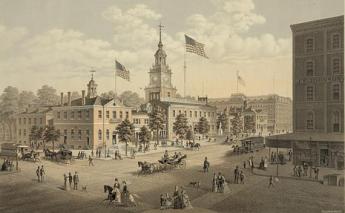
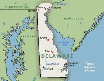 Originally the "lower counties" of Pennsylvania, and thus one of three Quaker colonies founded by William Penn, Delaware has developed its own set of traditions and history.
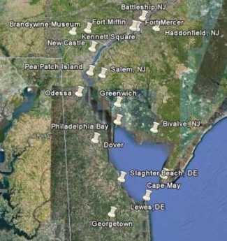 Start in Philadelphia, take two days to tour around Delaware Bay. Down the New Jersey side to Cape May, ferry over to Lewes, tour up to Dover and New Castle, visit Winterthur, Longwood Gardens, Brandywine Battlefield and art museum, then back to Philadelphia. Try it!
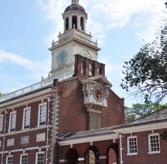 Millions of eye patients have been asked to read the passage from Franklin's autobiography, "I walked up Market Street, etc." which is commonly printed on eye-test cards. Here's your chance to do it.
 In 1751, the Pennsylvania Hospital at 8th and Spruce was 'way out in the country. Now it is in the center of a city, but the area still remains dominated by medical institutions.
 Grievances provoking the American Revolutionary War left many Philadelphians unprovoked. Loyalists often fled to Canada, especially Kingston, Ontario. Decades later the flow of dissidents reversed, Canadian anti-royalists taking refuge south of the border.